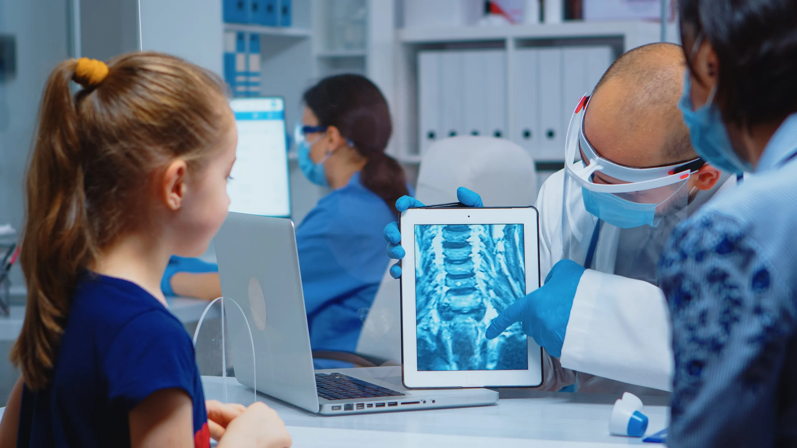
Have you ever wondered how doctors can see what’s inside your body without making any cuts? Well, that’s where X-rays come in! These tools have revolutionised medical diagnostics for over a century, allowing healthcare professionals to see inside the human body without surgery. Today, we’re diving deep into general X-rays, exploring everything from how they work to their benefits and potential risks.
What is a General X-ray?
A general X-ray is a quick, painless, and the most basic form of imaging test that uses small amounts of radiation to create pictures of the inside of your body. It’s like taking a photo, but instead of capturing what’s on the surface, it reveals what’s hiding beneath your skin and muscles.
How Does It Work?
Imagine you’re playing a game of shadow puppets. When you shine a light through your hands, it creates a shadow on the wall, right? X-rays work on a similar principle, but instead of visible light, they use X-ray radiation to produce images. This radiation passes through your body, and different tissues absorb it at different rates. Bones, being dense, absorb more radiation and appear white on the image, while softer tissues let more radiation pass through and appear darker. This technology helps doctors diagnose a wide range of medical conditions.
Common Uses of General X-Rays
Diagnosing Fractures and Bone Issues
X-rays are a common diagnostic tool for detecting bone fractures or dislocations. By clearly showing the position and condition of the bones, they assist in selecting the best treatment option, such as casting or surgery.
Detecting Infections or Lung Conditions
X-rays can detect lung infections, like pneumonia, and monitor conditions like asthma or chronic bronchitis. Just by visualising the lungs, doctors can assess the severity of the condition.
Screening for Tumours or Cancer
X-rays can sometimes be used to detect abnormalities in soft tissues, such as tumours. While they may not always provide detailed images of soft tissue, they are often the first line of screening before more advanced imaging methods.
The General X-Ray Procedure
- Preparing for the X-Ray
- Preparation for an X-ray is usually minimal. However, depending on the area being examined, you might be asked to:
- Remove jewellery or metal objects
- Change into a hospital gown
- Avoid eating or drinking for a few hours before the exam (in some cases)
- You may be asked to wear a lead apron to protect other parts of your body (in some cases).
What Happens During the Procedure?
During the X-ray procedure, a technician will position you, and the X-ray machine will be directed towards the area of concern. The procedure is rapid and painless, generally lasting only a few minutes. To avoid blurry images, you may be asked to hold your breath for a short time.
What to Expect After the X-Ray?
Getting the Results
Once the X-ray is finished, a radiologist will examine the images, interpret the findings, and communicate the results to your doctor. Typically, this process takes a day or two, but in emergencies, results can be provided more quickly.
Follow-up Steps If Necessary
If any abnormalities are detected, your doctor might recommend additional tests or treatments based on the findings. In some cases, more imaging, like an MRI or CT scan, might be required for a clearer view.
Types of X-ray Examinations
Chest X-ray
This is the most common type of X-ray. It’s like taking a snapshot of your heart, lungs, and chest bones. Doctors use it to check for pneumonia, heart problems, and even lung cancer.
Bone X-ray
Got a suspicion that you might have broken a bone? A bone X-ray is your go-to exam. It’s perfect for spotting fractures, dislocations, or even arthritis.
Abdominal X-ray
When your tummy’s giving you trouble, an abdominal X-ray can help pinpoint the issue. It’s great for detecting kidney stones, intestinal blockages, or swallowed objects.
Dental X-ray
Dental X-rays help dentists see what’s going on beneath your pearly whites, catching cavities and other dental issues before they become major problems.
Benefits of General X-rays
Non-invasive nature
One of the biggest benefits of X-rays is that they provide valuable insights into your health without surgery or invasive procedures. Most patients experience no discomfort or pain during the process.
Quick and convenient
The entire procedure, from positioning to taking the image, typically takes around 10-15 minutes, depending on the area being examined and the number of images needed. This rapid process makes it well-suited for emergencies or situations demanding a quick diagnosis, such as trauma cases or determining bone fractures.
Versatility in diagnosis
X-rays can help diagnose a wide range of conditions, from broken bones to lung diseases and beyond. This versatility of X-rays makes it a go-to diagnostic tool for doctors across many specialties.
Risks and Safety Considerations
Exposure to radiation
While X-rays are generally safe, they do expose you to a small amount of radiation. For most people, the benefits far outweigh the risks, but it’s important to limit unnecessary exposure.
Risk of Cancer
Excessive radiation exposure over time does carry a risk of causing cancer later in life, but the levels of radiation used in a typical X-ray are considered very low. Technicians use the lowest dose possible to achieve clear images.
Contrast medium reactions
Sometimes, to get a clearer picture, you might be asked to drink or receive an injection of a contrast medium. While reactions to these substances are rare, some people might experience allergic reactions or kidney problems. But don’t worry – medical staff are well-prepared to handle any issues that might arise.
Minimising Risks in X-Ray Procedures
ALARA (As Low As Reasonably Achievable) Principle
Healthcare professionals adhere to the ALARA principle, which means using the lowest possible radiation dose necessary to achieve a diagnostic image.
Technological Advancements
- Digital X-rays: The widespread adoption of digital X-ray technology has reduced radiation exposure compared to traditional film-based X-rays.
- Collimation: Collimators are used to restrict the X-ray beam to the area of interest, minimising unnecessary exposure.
- Grids: Grids are placed between the patient and the X-ray detector to absorb scattered radiation, improving image quality while reducing patient exposure.
Protective Gear and Shields
Patients may be given lead aprons or shields to protect specific areas of the body from unnecessary radiation exposure, especially during dental or abdominal X-rays.
Who Should Avoid X-Rays?
Pregnant Women
Pregnant women are advised to avoid X-rays unless absolutely necessary, as the radiation can affect the developing foetus. Your doctor might suggest alternative imaging methods or take extra precautions to protect your little one.
People with Certain Health Conditions
In some cases, individuals with specific health conditions, like those with implanted medical devices, might need to take special precautions or avoid certain types of X-rays.
Comparing X-rays to Other Imaging Techniques
CT scans
CT scans combine multiple X-ray images from different angles to create detailed cross-sectional views of the body. While they provide more information, they also use more radiation than general X-rays.
MRI
MRI, or Magnetic Resonance Imaging, uses powerful magnets and radio waves instead of radiation. It provides more detailed images of soft tissues than X-rays but takes longer and can be noisy.
Ultrasound
Using sound waves to create images, ultrasound is radiation-free and great for looking at soft tissues and blood flow. However, it can’t see through bone or air-filled spaces as X-rays can.
Radiology Imaging Solutions
At Radiology Imaging Solutions, we provide diagnostic imaging services to help doctors diagnose and treat a wide range of health conditions. Whether you need a general X-ray appointment or a more advanced imaging service, our expert team ensures you receive the highest quality care in a comfortable setting. Book your X ray appointment today and experience fast, reliable diagnostic services you can trust.


Leave a Reply